

Applied Physics | Bioscience | Computer Science | Material Science and Engineering
Colorful solution to advanced disease diagnosis
Nanophotonic microscope slides spotlight cancer cells hiding in patient tissue samples.


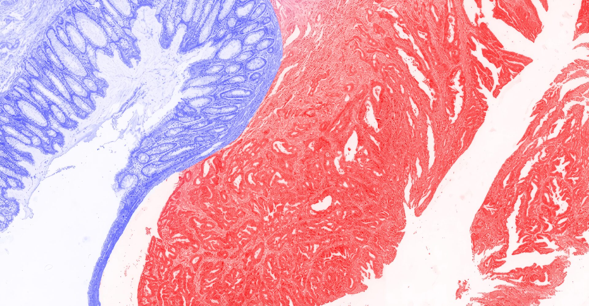
Cancer patients could gain accelerated access to potentially life-saving treatments, following the development of a high-tech, low-cost nanophotonic platform that aims to streamline patient tissue sample analysis.
In suspected cases of cancer and other diseases, diagnosis is confirmed by highly trained pathologists analyzing patient tissue samples under the microscope. To enhance the color contrast between different cell structures and tissue types, chemical dyes are added, making the telltale signs of cancer and other health conditions easier to spot.
Staining tissue samples for disease diagnosis requires specialist skill. However, it is costly, time-consuming, and prone to inconsistencies. KAUST’s Qiaoqiang Gan and his collaborators therefore set out to develop an alternative approach.
“Our goal was to develop a stain-free optical microscopy platform that could still provide pathologically meaningful cellular and tissue-level information,” says Qizhe Chen, a student in Gan’s lab.
Rather than relying on chemical dyes, Gan and his team focused on “structural color” to enhance the cellular features that pathologists use to diagnose disease[1]. Structural color arises in materials with nanoscale surface structures so fine that they interact with light, absorbing some wavelengths while reflecting others to create color.
Butterfly wings and peacock feathers are natural materials with brilliant structural color. It can also be designed into synthetic materials with nanostructured surfaces, says Gan, who has studied nanophotonics and structural color for nearly two decades.
“Since joining KAUST, I have been interested in applying these principles to real-world problems,” Gan says. “Through discussions with our clinical collaborators at Huashan Hospital in Shanghai, we realized that structural color could potentially provide a new contrast mechanism for tissue imaging without relying on traditional chemical staining.”
The team designed structurally colored microscope slides made from silicon wafers nanocoated with silicon nitride. “The silicon nitride nanolayer acts as an ‘optical nanocavity’,” Chen says. When light reflects off the slide, the nanocoating selectively enhances certain wavelengths while suppressing others. The team harnessed this tuned reflectivity to amplify optical differences between cancerous and healthy tissue. “The slides display these differences as distinct colors, making cancer-associated regions easier to distinguish from healthy tissue,” Chen says.
The team used real tissue samples from colorectal cancer patients to demonstrate the diagnostic potential of their nanocavities-on-silicon (NOS) slides, says Yanyan Li, a postdoc in KAUST’s Bioscience program who collaborated with Gan’s team. “The slides provided sufficient color contrast to visualize differences between normal and cancerous colorectal tissue regions without conventional staining,” she says. An additional advantage is that, by preserving the tissue sample in an unstained state, pathologists could run additional cancer assays on the same slide, further streamlining the diagnostic workflow, Li notes.
NOS slides are simple and inexpensive to make, which is crucial for high-throughput biomedical use. The team was able to record thousands of NOS slide microscope images of healthy and cancerous tissue – capturing enough image data to train an AI model to automatically detect colorectal cancer in NOS microscope images.
“The model showed strong performance in distinguishing healthy from cancerous colorectal tissue, achieving greater than 95 percent accuracy,” says Lijie Hu, a student in the Computer Science program who collaborated on the project.
Gan and his team are now exploring whether nanophotonic technology could reveal additional information in tissue samples, for advanced disease diagnostics beyond conventional staining. They are also building large, high-quality NOS imaging datasets for AI-assisted diagnosis, Gan adds.
“We are working with colleagues at KAUST, and at local hospitals, to implement and validate this technology in clinically relevant settings,” Gan says. “Our long-term goal is to combine nanophotonic stain-free imaging with AI to address important healthcare challenges in Saudi Arabia and beyond, making pathology faster, more scalable, and potentially more accessible.”
Reference
- Chen, Q., Ren, Y., Hu, L., Li, Y. Liang, W., Wang, J., Gao, H., Wang, X., Li, J., He, Q., Zhu, Y., Hu, H., Zhan, Q., Gallouzi, I., Merzaban, J., Wang, D., Du, Z., Gu, X., Gan, Q. Intelligent stain-free histology on structural colorimetric nanocavities. Advanced Science 13, e14340 (2026).| article.
You might also like

Computer Science
Virtual reality headsets open a window into the mind
Bioscience
Multi-enzyme pathway delivered into living cells
Bioscience
Brain fuel also helps wire neural connections
Bioscience
Single small-molecule model reveals insights into human embryo development

Bioscience
Can AI finally bring order to biology’s data deluge?

Computer Science
AI-based hydrogen plant models improve power grid stability
Bioengineering
Bio-inspired network structures for next-generation AI
Bioscience