Chemistry
High-sensitivity cameras reveal the atomic structure of metal-organic frameworks
Highly sensitive electron cameras allow researchers to see the atomic structure of metal-organic frameworks.


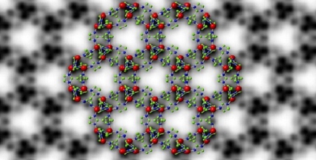
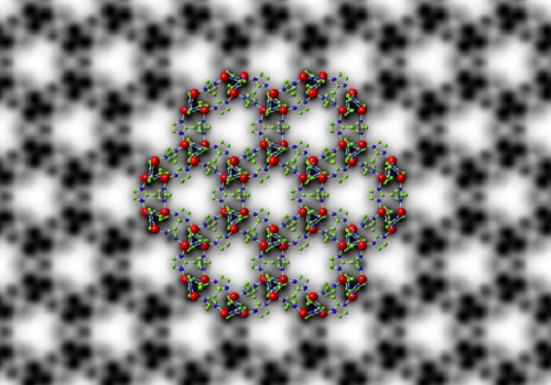
Symmetry-imposed and lattice-averaged HRTEM image of the metal–organic framework ZIF-8 (black and white) with a structural model overlaid to show the position of the zinc ions and organic ligands (in color).
© 2017 KAUST
A method for fine-scale imaging of a class of materials that is useful for gas storage and separation is needed. However, typical visualization methods emit an electron dose that can cause damage to the structure of some materials.
One of these materials are metal-organic frameworks (MOFs), three-dimensional structures made up of metal ions connected by organic ligands. MOFs are useful for gas storage and separation because they can be designed to have precise pore sizes of molecular dimensions and large void spaces (porosity) within their frameworks.
“To thoroughly understand the performance of metal-organic frameworks in various applications, we need to know their structures at the atomic level because their macroscopic behavior is determined by their microscopic structure,” explained Yu Han, KAUST professor of chemical science. By visualizing these structures, researchers can uncover important clues about how these materials self-assemble to create their trademark pores.
Several members of the University’s Advanced Membranes and Porous Materials Center joined forces with the University’s Imaging and Characterization Core Lab and with colleagues from Gatan, a company specializing in electron microscopy, Lawrence Berkeley National Laboratory and others in China. Their collaboration resulted in an adaptation of high-resolution transmission electron microscopy (HRTEM) using state-of-the-art direct-detection electron-counting cameras.
The downfall of using HRTEM to image MOFs is that the high-energy electrons that pass through the sample cause damage to the structure. By modifying HRTEM using high-sensitivity detectors, the team acquired images with an electron dose low enough not to damage the structure of MOFs. Ultimately, the group produced high-resolution images of their atomic structures.
As a study case, the team applied their method to ZIF-8, a MOF comprising zinc ions connected by organic 2-methylimidazole linkers. They were able to image its structure with a resolution of 0.21 nanometers (one nanometer is one billionth of a meter), a resolution high enough to image the individual columns of zinc atoms and organic linkers.
This helped the researchers to reveal the surface and interfacial structures of ZIF-8 crystals. “The results unraveled that porosity generated at the interfaces of ZIF-8 crystals is different from the intrinsic porosity of ZIF-8, which influences how gas molecules transport in ZIF-8 crystals,” explained Han.
This work sets the foundation for the use of non-destructive atomic-level HRTEM imaging of other materials, including polymers and two-dimensional materials.
References
- Zhu, Y., Ciston, J., Zheng, B., … & Han, Y. Unravelling surface and interfacial structures of a metal–organic framework by transmission electron microscopy. Nature Materials 16 532-536 (2017).| article
You might also like

Chemistry
Opening a window on architectural nano-design
Bioscience
Multi-enzyme pathway delivered into living cells

Chemical Engineering
Natural solvents enable fully bio-based membranes

Chemistry
Turning infrared solar photons into hydrogen fuel

Applied Physics
Natural polymer boosts solar cells
Chemistry
Disruptive smart materials flex with real world potential
Chemistry
Catalysts provide the right pathway to green energy
Chemistry