Bioscience
Ultrabright dots see beyond skin deep
Tiny light-emitting probes give researchers a better option for noninvasive imaging of living tissue.


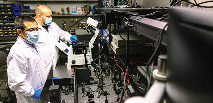
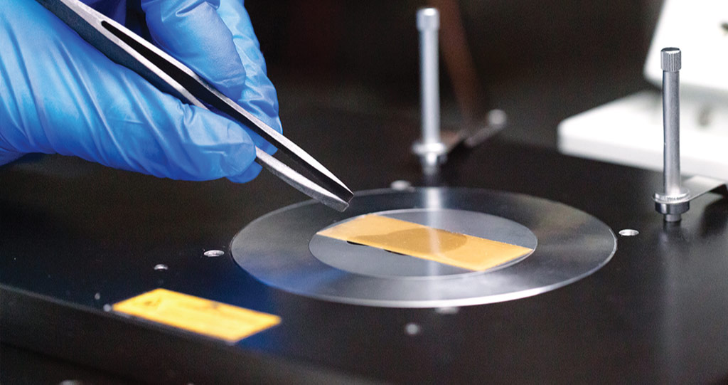
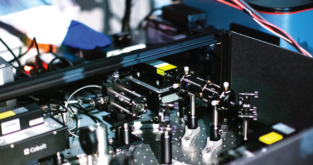
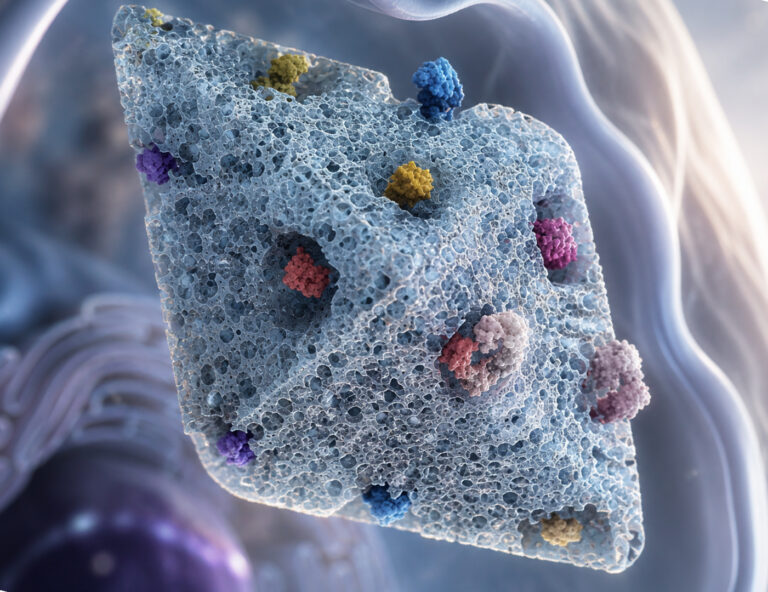
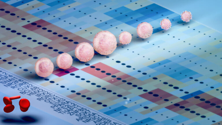
KAUST researchers have developed a custom-designed polymer to produce light that penetrates murky environments, such as inner organs. The light-emitting material has shown promise in bioimaging trials. © 2021 KAUST; Anastasia Serin.
KAUST researchers have developed a custom-designed polymer to produce light that penetrates murky environments, such as inner organs. The light-emitting material has shown promise in bioimaging trials. © 2021 KAUST; Anastasia Serin.
A polymer that is custom designed to produce light that penetrates murky environments has shown promise in bioimaging trials, where it can detect nano-sized particles underneath the surface of realistic tissue models.
Recent studies have demonstrated that fluorescent probes, which are light-emitting materials that attach to tiny targets such as cells, are particularly useful for bioimaging when they radiate in the shortwave infrared (SWIR) region of the optical spectrum. Because this type of fluorescent light penetrates deeper into biological objects without being absorbed or scattered, SWIR probes can be spotted farther into tissue than conventional emitters. These features have enabled SWIR probes to capture high-resolution images of structures located deep within the body, such as brain tissue, without the hazards of x-rays.
Polymers offer an alternative to semiconductor quantum dots or rare-earth-doped nanoparticles that are unsuitable for many specimens because of their toxic side effects.
© 2021 KAUST; Anastasia Serin
Satoshi Habuchi and his colleagues are working to improve fluorescent imaging by expanding the type of probes capable of producing SWIR radiation. Currently, most bright SWIR emitters are either semiconductor quantum dots or rare-earth-doped nanoparticles that are unsuitable for many specimens because of their toxic side effects. On the other hand, materials that are more biocompatible, such as organic dyes, are usually not intense enough to be seen inside tissue.
To resolve this issue, KAUST researchers turned to polymers having “donor–acceptor” structures, a layout where electron-rich components alternate with electron-poor portions along a conductive molecular chain. “This distribution promotes charge transfer along the polymer backbone, which is a very effective way to obtain SWIR light,” explains Hubert Piwoński, the study’s lead author.
“Our polymer dots are a big step toward single-particle tissue imaging.”
The team chose two donor–acceptor polymers with ideal characteristics for SWIR emission and then developed a precipitation procedure that fused the compounds into tiny polymer spheres, or “dots”, just a few nanometers wide. Optical characterizations revealed these materials had exceptionally bright SWIR emissions that were easily spotted in biological tissue models.
The team’s particles are so bright that they enable detection of nanometer-sized polymer dots in specimens one millimeter thick.
© 2021 KAUST; Anastasia Serin
“Per volume, our particles have a brightness value larger than almost all other SWIR emitters reported so far,” says Habuchi. “This enabled detection of nanometer-sized polymer dots in specimens one millimeter thick.”
In addition, the new polymer dots that fluoresce only for a nanosecond can produce low-noise images with single-molecule sensitivity due to high throughput detection of emitted fluorescence. The ability to visualize single probes at fast acquisition rates could benefit researchers looking to capture processes in tissues and organs as they happen.
“There are huge opportunities for new probes and imaging modalities capable of addressing the dynamics of molecules in living systems, and our polymer dots are a big step toward single-particle tissue imaging,” says Piwoński.
References
- Piwoński, H., Wang, Y., Li, W., Michinobu, T. & Habuchi, S. Millimeter-deep detection of single shortwave-infrared-emitting polymer dots through turbid media. Nano Letters 20, 8803–8810 (2020).| article
ABOUT THE AUTHOR
Hubert Piwoński
Research Scientist

You might also like
Applied Physics
Colorful solution to advanced disease diagnosis
Bioscience
Multi-enzyme pathway delivered into living cells
Bioscience
Brain fuel also helps wire neural connections
Bioscience
Single small-molecule model reveals insights into human embryo development

Bioscience
Can AI finally bring order to biology’s data deluge?
Bioengineering
Bio-inspired network structures for next-generation AI

Bioscience
Robust workflow built for chemical genomic screening
Bioscience