Bioengineering
Ultrashort peptides go a long way for tissue engineering
A printable hydrogel made of ultrashort peptides could help shape cells into viable tissues.


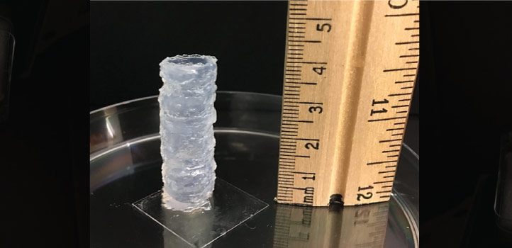
KAUST bioengineers have developed a bioprinting process that uses ultrashort peptides as the basis of the scaffolding ink. © 2021 KAUST. /en/article/1112/ultrashort-peptides-go-a-long-way-for-tissue-engineering
KAUST bioengineers have developed a bioprinting process that uses ultrashort peptides as the basis of the scaffolding ink. © 2021 KAUST. /en/article/1112/ultrashort-peptides-go-a-long-way-for-tissue-engineering
A new automated process prints a peptide-based hydrogel scaffold containing uniformly distributed cells. The scaffolds hold their shapes well and successfully facilitate cell growth that lasts for weeks.
“Bioprinting” — 3D printing that incorporates living cells — has the potential to revolutionize tissue engineering and regenerative medicine. Scientists have experimented with natural and synthetic “bioinks” to print out scaffolds that hold cells in place as they grow and form a tissue with a specific shape. But there are challenges with cell survival. Natural bioinks, such as gelatin and collagen, need to be treated with chemicals or ultraviolet light to hold their shape, which affects the cell viability. The synthetic polymer hydrogels tested to date also require the use of harsh chemicals and conditions that threaten cell survival.
KAUST bioengineer Charlotte Hauser led a team of researchers to develop a bioprinting process that uses ultrashort peptides as the basis of the scaffolding ink. They designed three peptides using different combinations of the amino acids isoleucine, lysine, phenylalanine and cyclohexylalanine.
For the actual printing, the team used a novel triple-inlet nozzle. The peptide bioink goes into one inlet, a buffer solution goes into another, and cells are added through a third. This allows the peptide ink to gradually mix with the buffer solution and then combine with the cells at the nozzle’s outlet. Once the ink is ejected, it instantly solidifies, capturing the cells within its structure.
“It’s challenging to find a cell-friendly biomaterial that supports long-term cell survival and is also printable,” says Ph.D. student Hepi Hari Susapto. “Our bioinks made from self-assembling ultrashort peptide hydrogels efficiently address this challenge.”
The team was able to print cylinders up to four centimeters tall, such as in the image above, and a human-like nose, which all held their shapes well.
Human fibroblasts, human bone marrow mesenchymal stem cells and mouse brain neurons survived and proliferated well within the hydrogel matrix. The scientists further induced bone marrow mesenchymal stem cells to differentiate inside a printed scaffold into elastic cartilage-like tissue within a period of four weeks.
The team is now working on changing the surface chemistry of their bioinks so that they more closely resemble the cell environment in the human body.
“Our next step is to bioprint 3D disease models and miniature organs for high-throughput drug screening and diagnosis,” says Hauser. “These could help reduce the time and cost of searching for more effective and personalized drugs.”
References
- Susapto, H.H., Alhattab, D., Abdelrahman, S., Khan, Z., Alshehri, S., Kahin, K., Ge, R., Moretti, M., Emwas, A. & Hauser, C.A.E. Ultrashort peptide bioinks support automated printing of large-scale constructs assuring long-term survival of printed tissue constructs. Nano Letters 7, 2719–2729 (2021).| article
ABOUT THE AUTHOR
Hepi Hari Susapto
Ph.D. Student

You might also like
Bioengineering
Smaller and sharper: Compact CRISPR scissors cut with precision
Bioengineering
Intelligent vision chip offers brainlike processing
Bioengineering
Bio-inspired network structures for next-generation AI
Bioengineering
Self-aware biosensors boost digital health monitoring
Bioengineering
Algae and the chocolate factory
Bioengineering
Smart patch detects allergies before symptoms strike
Bioengineering
Cancer’s hidden sugar code opens diagnostic opportunities
Bioengineering