Bioscience
Finding a handle to bag the right proteins
A method that lights up tags attached to selected proteins can help to purify the proteins from a mixed protein pool.
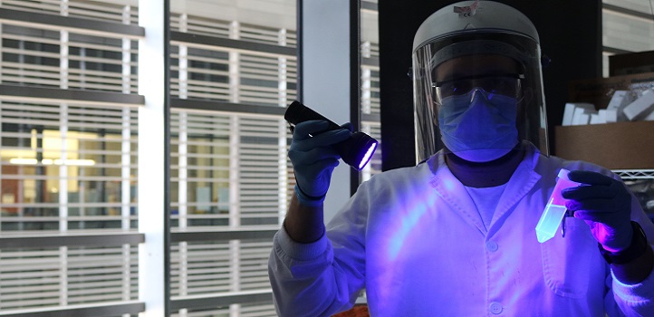
Purifying specific protein molecules from complex mixtures will become easier with a simpler way to detect a molecular “tag” commonly used as a handle to grab the proteins.
Proteins, comprising many linked amino acid molecules, form the key “workforce” of molecular biology, performing a multitude of chemical tasks, including catalyzing the chemistry of life, switching genes on and off, and receiving and responding to signals between cells.
Researchers need to produce and purify selected proteins to investigate their activities for drug research, biotechnology and basic investigations of cell biology.
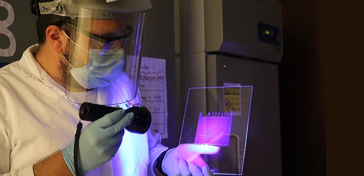
When the gel is stained with the conjugate and illuminated with UV radiation, the His-tagged proteins can be seen with the naked eye.
© 2020 KAUST
Proteins of interest are commonly made by inserting the genes that code for them into cells that will produce them, but that leaves the problem of identifying and purifying the desired protein from a potentially complex mixture. A common strategy is to modify the gene encoding the protein to make the protein carry a string of molecules of the amino acid called histidine, creating a “polyhistidine tag.”
“The tag acts like a handle attached to a bag,” explains the first author of the study, Vlad-Stefan Raducanu. “It’s much easier to fish out a protein by catching the tag.”
The various proteins in an impure sample can be separated using an electric field to pull them through a gel at different rates—a process called gel electrophoresis. The gel is then transferred to a membrane and the region carrying the polyhistidine-tagged proteins is visualized using antibodies, also a form of protein, to selectively bind to the tag. However, this type of detection can be laborious.
Now, Raducanu and his colleagues have developed a simpler detection procedure that avoids the membrane transfer step and the use of antibodies.
They constructed a chemical complex that binds to polyhistidine tags and can be stimulated by ultraviolet (UV) radiation to fluoresce with visible light. The regions of the gel-carrying tagged proteins can be readily detected by the light given off by the UV-excited “fluorophore” complexes bound to the tags.
“It was challenging to devise a suitable UV-excitable fluorophore,” Raducanu explains. The team had to couple the fluorescent component of their complex to another part containing a metal ion that can bind to the polyhistidine tag.
“We now plan to collaborate with chemists at KAUST to develop even brighter dyes,” Raducanu says, expressing hope that the usefulness of UV-excitable fluorophores could be adopted more widely to help researchers detect the proteins they need.
References
- Raducanu, V-S., Isaioglou, I., Raducanu, D-V., Merzaban, J.S. & Hamdan, S.M. Simplified detection of polyhistidine-tagged proteins in gels and membranes using a UV-excitable dye and a multiple chelator head pair. Journal of Biological Chemistry 295, 12214-12223 (2020).| article
You might also like
Bioscience
Robust workflow built for chemical genomic screening
Bioscience
Cell atlas offers clues to how childhood leukemia takes hold
Bioscience
Hidden flexibility in plant communication revealed
Bioscience
Harnessing the unintended epigenetic side effects of genome editing
Bioscience
Mica enables simpler, sharper, and deeper single-particle tracking
Bioengineering
Cancer’s hidden sugar code opens diagnostic opportunities
Bioscience
AI speeds up human embryo model research
Bioscience


