Bioscience
The brain gamers
Novel use of gaming technology enables researchers to 'step inside' the brain.


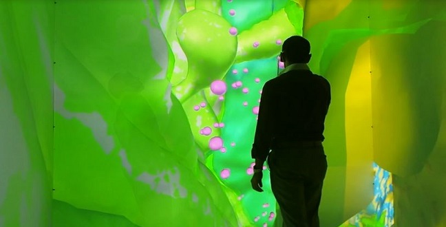
Technology that creates a 3D virtual reality is best known to gamers, and it has now given researchers new insight into the structure of the brain, enabling them to inspect specific neural structures with unprecedented clarity.
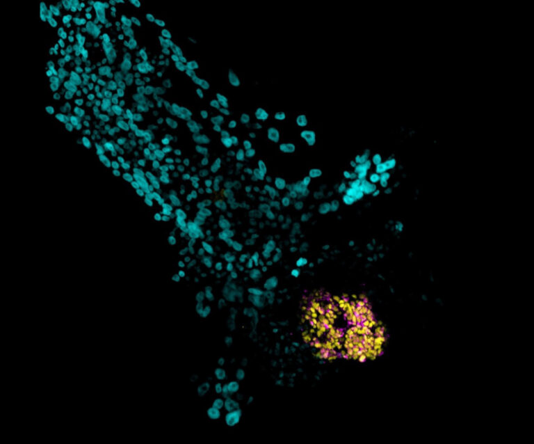
Photographs of cross-sections of mouse brain tissue scanned under an electron microscope were reconstructed using special software, allowing a multidisciplinary team of researchers to examine the tissue in a 3D virtual environment1.
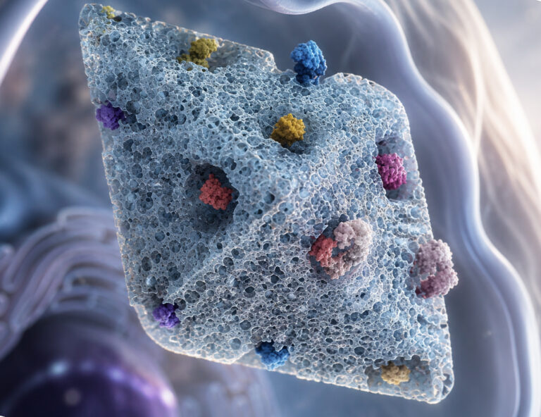
“Aside from the excitement of being able to physically enter a 3D microscopic model of the brain, we can see new patterns in the neural tissues,” said neurobiologist Pierre Magistretti from KAUST’s Biological and Environmental Science and Engineering Division. “For instance, we could show that glycogen granules, a major source of energy in the brain, are not randomly located but accumulate around synapses, the sites of contact between neurons, which consume a lot of energy in order to work properly.”
Such a detailed study on the ultra-structural localization of glycogen in the brain is new.
“This is the first time that such a 3D virtual reality analysis of brain structure has been performed at such a high level of spatial resolution,” said Corrado Cali, a postdoctoral fellow in Magistretti’s lab and first author of the paper.
The research was developed by KAUST biological researchers, the University’s Visualization Lab, Germany’s Heidelberg Collaboratory for Image Processing and Switzerland’s Brain Mind Institute in Lausanne.
Researchers step into a three-meter cubical room that has projectors on all six sides. Using special 3D glasses, they gain the illusion of standing within the projected 3D image of the brain tissue. The high resolution of the images allowed them to identify and precisely locate various neural tissues as well as the locations of minute subcellular features such as glycogen granules.
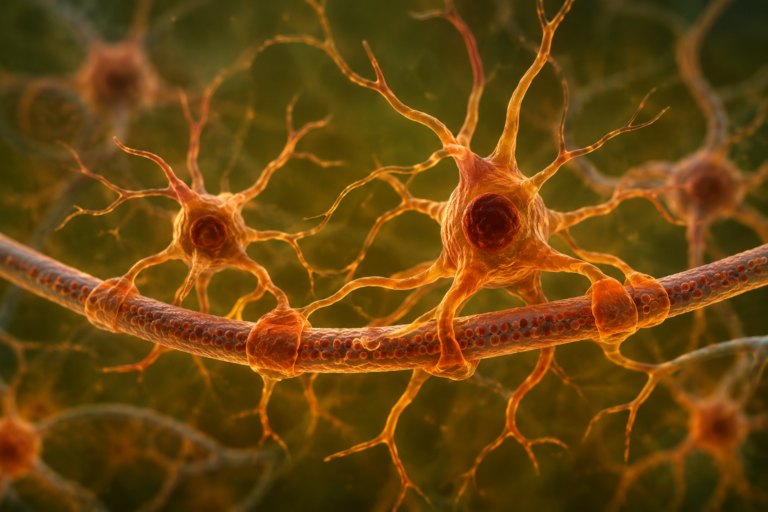
The team found that a significant portion of glycogen granules collected around neuron synapses close to the walls of blood vessels. This suggests that glucose entering the brain parenchyma from the blood stream is immediately converted to glycogen.
Glycogen breaks down in the brain to produce lactate, which is then shuttled to neurons, playing a vital role in learning and memory.
“The result was beyond our expectations,” said Magistretti. “The tool is extremely useful to help us to investigate, brainstorm and decide how to modify our analysis tool to improve our observations.”
The team plans to use the same approach to study how memory is affected in animals following disturbances of glycogen metabolism. They also plan to use 3D virtual reality environments to examine pathological brain samples, with particular emphasis on epilepsy and neurodegenerative diseases.
References
- Cali, C., Baghbara, J., Boges, D. J., Holst, G. R., Kreshuk, A. et al. Three-dimensional immersive virtual reality for studying cellular compartments in 3D models from EM preparations of neural tissues. The Journal of Comparative Neurology 524, 23-38. | article
You might also like
Applied Physics
Colorful solution to advanced disease diagnosis
Bioscience
Multi-enzyme pathway delivered into living cells
Bioscience
Brain fuel also helps wire neural connections
Bioscience
Single small-molecule model reveals insights into human embryo development
Bioscience
Can AI finally bring order to biology’s data deluge?
Bioengineering
Bio-inspired network structures for next-generation AI

Bioscience
Robust workflow built for chemical genomic screening
Bioscience