Statistics
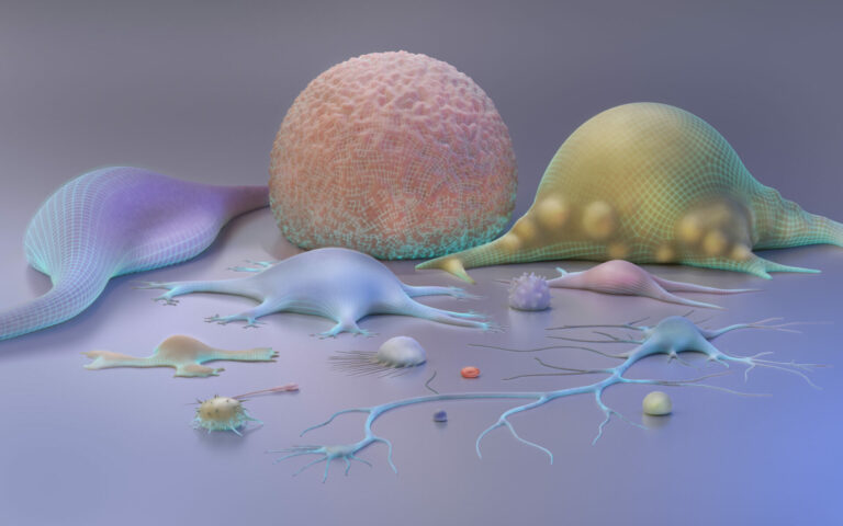
Zooming in on brain function
Faster computations will allow researchers to see the finer details of brain activity in functional brain imaging.


A computationally efficient data-processing scheme will make it possible to see correlations in brain activity between different parts of the brain at unprecedented resolution. The statistics-based computation scheme, developed by KAUST researchers, also tackles one of the most critical problems of medical and biological imaging—how to process imaging data fast enough to realize the full exploratory power of the latest high-resolution imaging techniques.
The development of functional magnetic resonance imaging (fMRI) in the early 1990s was one of those true Eureka moments for brain research. Using existing noninvasive MRI technology, fMRI maps the distribution of blood oxygen in the brain, which is closely correlated with brain activity. With fMRI, it is possible to take snapshots of brain activity in response to specific stimuli, such as speech, responding to memory questions or visual scenes.
While fMRI is capable of taking high-resolution images consisting of hundreds of thousands of points or voxels, it is an enormous computational task to map the correlations between simultaneous brain function across different areas of the brain. Even with the range of computing power available today, such computations are not feasible by direct methods and so require a more computationally efficient approach.
Marc Genton and Hernando Ombao from KAUST, in collaboration with Stefano Castruccio from the University of Notre Dame in the United States, have addressed this problem. They developed a statistics-based computational scheme that matches activity in different parts of the brain at different spatial scales, from whole-of-brain to smaller regional structures and down to the tiniest brain volume.
“Using a statistical approach, our multiresolution approach essentially breaks up the spatial component of the fMRI data into different scales—from global to local,” says Ombao.
Developed as part of a collaboration with the stroke rehabilitation center at the University of California Irvine, the computation scheme calculates the statistical shape of increasingly large populations of activity readings in a way that lends itself readily to distributed computing, making it highly efficient.
Not only does this solve the computational issue, it improves the interpretability of results, ensuring that the connectivity between activities can be characterized both within each brain region and across different brain regions.
“It is important to take into account how spatially separated neuronal units communicate with each other in modeling fMRI data in order to avoid misleading results, such as false activations, or an inability to detect activity.” says Ombao. “Correct identification of activated and inactivated units will help us to improve our understanding of human brain function in both healthy and diseased populations.”
References
-
Castruccio, S., Ombao, H. & Genton, M.G. A scalable multi-resolution spatio-temporal model for brain activation in fMRI. Biometrics early online article 22 January 2018.| article
ABOUT THE AUTHOR
Statistics Program

You might also like

Statistics
Checking your assumptions

Statistics
Internet searches offer early warnings of disease outbreaks

Statistics
Joining the dots for better health surveillance

Statistics
Easing the generation and storage of climate data

Statistics
A high-resolution boost for global climate modeling

Applied Mathematics and Computational Sciences
Finer forecasting to improve public health planning

Bioengineering
Shuffling the deck for privacy
Bioengineering