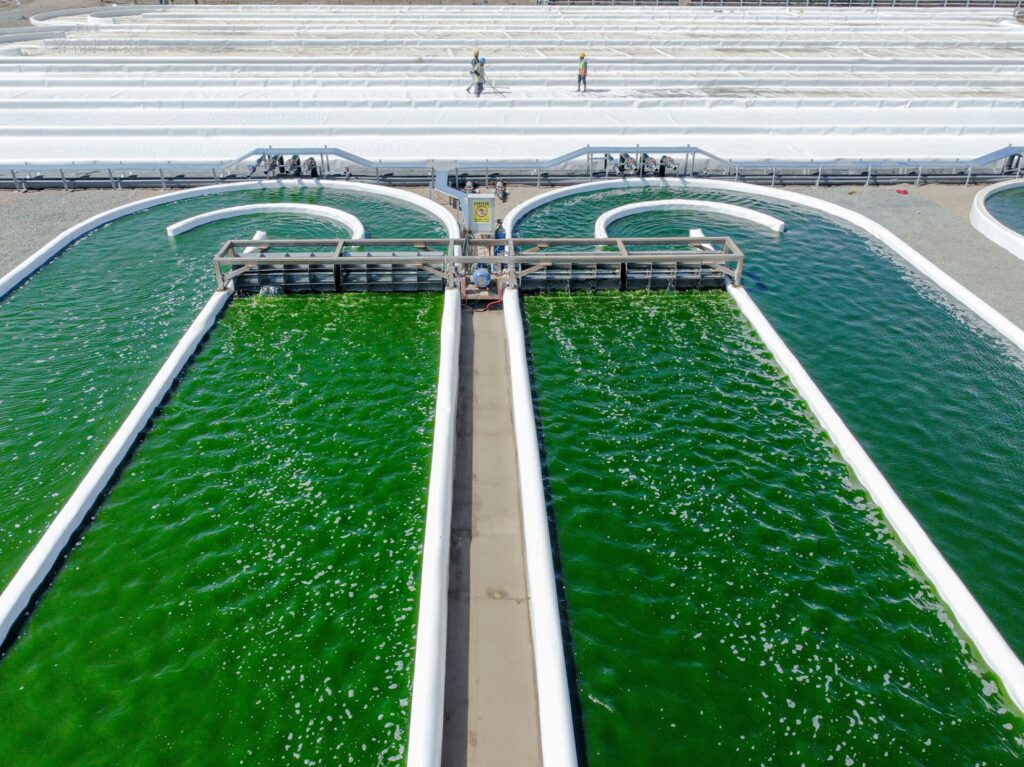
A greener future for Saudi Arabia and beyond may emerge from an unexpected source: algae. Over the past five years, KAUST researchers have laid the groundwork for a new industry in the Kingdom. Their work spans the cultivation, harvesting, and processing of microalgae and macroalgae as sources of sustainable animal feed, biomolecules for medicine, carbon dioxide conversion, and raw materials for textile production for the fashion industry.
“Algae are among the most flexible organisms on Earth,” says Claudio Grünewald, who leads algal research at KAUST. “Algae are key photosynthetic organisms that uptake, utilize and sequester carbon dioxide, and produce copious oxygen. We have a fantastic opportunity to harness the green potential of this highly adaptable organism.”
Grünewald, as project director at KAUST Beacon Development, oversees the advancement of groundbreaking algal technologies and techniques for successful farming of microalgae, and for the processing of wild macroalgae (seaweeds) found along the Red Sea coast.
With microalgae, the biomass generated can be used for industrial and agricultural applications. Macroalgae, meanwhile, are a valuable source of alginate, a polysaccharide used as a thickening and gelling agent. High-quality alginate is widely used in the food industry, while lower-quality grades are used as paint thickeners and in other industrial applications.
Algae cultivation potential in Saudi Arabia
“Saudi Arabia’s landscape and climate offer favourable growth conditions for algal farming,” says Grünewald. “We have the key parameters needed to grow microalgae at scale in semi-controlled environments: flat open spaces, high light radiation, pristine seawater, and sources of excess carbon dioxide.”
Microalgae use carbon dioxide as a main carbon source for photosynthesis that, in turn, generates oxygen, biomass and other valuable compounds. This natural potential to turn emissions into a useful resource opens the possibility for every thermal power plant to incorporate an algal farm as a carbon capture and conversion unit, supporting Saudi Arabia’s target of carbon neutrality by 2060. This vision is being driven by a growing partnership between KAUST and a major multinational company.
Grünewald’s team have built microalgae and seaweed processing facilities at KAUST and also with NAQUA, one of Saudi Arabia’s largest aquaculture companies. They have grown multiple species of microalgae in a wide variety of water types, from fresh water to highly saline brines from desalination plants, and proved that the resulting biomass is quality feed for chickens and fish. Moreover, these organisms can serve as a matrix for plant biostimulant production.
“We’ve tested microalgae bio-stimulants in several crops, including cherry tomatoes and potatoes, and results show that they can improve yields by 30 percent,” says Grünewald. Byproducts from seaweed processing can also be used as fertilizers to improve crop yields.
Solid support means that algal farms are spreading across the Kingdom. Since KAUST first began pioneering algal production techniques and technologies in 2021, at least six companies have either established or are in the process of establishing microalgae farms in the country.
“One of our main funders was the Ministry of Environment, Water and Agriculture,” says Grünewald. “We’ve validated our findings under strict environmental guidelines and demonstrated to investors that it is feasible to produce algae biomass at scale here. Both local and international companies are investing in the industry, and we expect algal farming to cover over 300 hectares by the end of 2028.”
All companies must adhere to environmental legislation for farming microalgae. Should any companies decide to grow species that are not native to the region, they must ensure proper treatment and secure growing facilities to avoid exotic species escape, notes Grünewald.
The algae industry will support national food security goals, providing a sustainable, local source of feedstock and fertilizers. It will create jobs and improve local economies by boosting the whole value chain, because the materials used to build and support these facilities are locally sourced.
Seaweed success stories
On the macroalgae side, Grünewald and co-workers have conducted extensive surveys into a brown seaweed called Sargassum, which is particularly prolific along the Red Sea coast. Sargassum flourishes from September to February, following a reproductive phase in late summer. The seaweed washes up in vast quantities along the coast; rotting seaweed is a health hazard, and acidic gases can also leach from the seaweed, degrading the environment and harming wildlife.
“By harvesting and processing Sargassum to produce alginate, there are many positive secondary effects,” says Grünewald. “We’ll employ local fishermen to harvest the seaweed during its peak season, providing incomes and taking the pressure off fisheries for part of the year. Local authorities will save money usually spent on cleaning up seaweed, and tourism will benefit from cleaner beaches. This seaweed also captures a lot of carbon dioxide from the atmosphere, and we can turn this into safe, usable forms of carbon.”
Alginate is currently imported from China in bulk by the food industry in the Kingdom. A secure local source of alginate will further improve food security and support local economies and employment. Before Sargassum processing is rolled out at scale, Grünewald is determined to ensure the harvest does not disrupt Red Sea ecosystems.
“Sargassum ‘forests’ normally grow in shallow water, and provide shelter and habitat to tiny organisms, including coral and fish larvae. We mustn’t disturb them,” says Grünewald. “With KAUST colleagues, we’re conducting research to understand the seasonal cycles of these creatures, and ensure we harvest the seaweed at the correct time each year.”
For now, KAUST is processing Sargassum in a pilot ‘biorefinery’, extracting alginate first, then using the leftover material to make other valuable products. This seaweed can be turned into fibers for making beautiful fabrics, for example.
“Both micro- and macro-algae are surprisingly versatile, and, alongside interested partners from all industries, we’ll continue to explore the uses stemming from algal processing,” says Grünewald.REFERENCES
Mhedhbi, E., Padri, M., Al Shaikhi, A., Al Hafedh, Y ., Fuentes Grunewald, C. The potential of microalgae to contribute to sustainable animal feed production in the Arabian Peninsula, Saudi Arabia. Applied Phycology7 (1), 2026. |article
KAUST converts Red Sea seaweed into economic and environmental value. |article
Silkina, A., Gayo-Pelaez, J.I., , Fernandes, F., Fuentes-Grünewald, C., Kapoore, R.V., Tang, K.W. From waste to wealth: coupling different nutritional modes of Scenedesmus obliquus for waste remediation and algal product development. Journal of Applied Phycology37 (2), 2025. |article
Over the last decade, research suggesting that forests can have warming impacts in certain regions of the globe sparked debate about their role in climate. Forest conservation and restoration projects have, at times, been paused over concerns that they could contribute to climate change rather than counteract it.
However, recent research takes a global perspective to help navigate past the controversy, showing forests’ vital roles in local climate adaptation. KAUST ecologist, Thomas Crowther, has led an international team that reviewed evidence and found that, when each forest climate impact study is considered in context, native forest protection and regeneration is overwhelmingly positive for humanity and the planet[1].
Location, location
“Our study was initiated by colleagues in the UK government who manage an international conservation and development budget,” says Crowther, whose research focuses on links between human wellbeing and global biodiversity. “The question over whether forests have a warming or a cooling effect was blocking their work.”
To resolve apparently conflicting research findings about forests’ impact on local climate, the team considered the locations the studies were conducted. “When we take that perspective, the controversy washes away and the evidence is crystal clear,” Crowther says.
The analysis showed that local warming was restricted to the snowy boreal forests of the Northern Hemisphere’s high latitudes. This local warming effect — which is beneficial for biodiversity and people in these ecosystems — stems from the way forests can absorb the warmth in sunlight, Crowther explains.
“By absorbing carbon, forests have a cooling impact, but in boreal forests, that effect can be offset by the warming of the Sun’s energy being absorbed,” Crowther notes. Compared to open, highly reflective snow-covered landscapes, dark forests absorb more solar energy and warm the local climate.
“We see a very clear pattern, particularly at the local scale, that forests in freezing conditions do have a local warming impact, which is beneficial to the people and species living there,” Crowther says.
“In hot locations, forests have an equally important cooling effect, saving lives and livelihoods in these regions in the face of climate change,” Crowther adds. “The amazing potential of forest ecosystems to buffer the effects of climate means that they’re beneficial wherever they grow naturally.”
Flow-on benefits
Forests’ local climate-regulating impacts extend beyond temperature. The team also weighed the evidence on water flows through forested landscapes.
“Forests slow down water cycling, which can trap moisture in the ecosystem and is very good for those ecosystems’ survival,” Crowther explains. “This effect can be really helpful in wet regions, where slowed water flows can prevent flooding,” he says.
However, he warned that the slowed water cycling can also be an important planning consideration in arid areas. “In dryland regions, living downhill from a forest can limit water runoff towards your community,” Crowther says.
For a country such as Saudi Arabia, the low moisture availability is a limiting factor in forest growth. “But there are huge areas of this country where trees naturally exist, for example, river basins and wadis with higher moisture availability,” Crowther says. “Those areas are incredible opportunities for forest regeneration, where buffering temperatures and retaining more moisture can be really valuable for local livelihoods and food production.”
Pinpointing the places where forests naturally grow is a major focus of Crowther’s research. “Our first model, published in 2019, was the basis for the United Nations’ Trillion Tree campaign, which inspired international forest restoration efforts including the Saudi Green Initiative,” Crowther explains. “Those original models have since been refined and we now can show accurately where forests would have enough access to moisture, and where reforestation would provide climate benefits.”
A world in balance
As Crowther’s models highlight, the natural world contains a diversity of ecosystems far beyond forests. Reforestation efforts must reflect this diversity, Crowther says. “Forest benefits are most consistent when they grow in places where they would naturally exist. When they are planted in unnatural areas, their effects can be highly unpredictable and often damaging.”
One place to focus forest conservation and restoration efforts is the hot tropics, where biodiversity, carbon storage and livelihoods depend most closely on natural forest ecosystems, Crowther points out. “At the same time, I firmly consider that there’s no location on the planet where healthy biodiversity isn’t beneficial for people,” he adds. “Where peatlands are naturally found, you want healthy peatland ecosystems there. In grassland regions, you want grassland biodiversity. I want a world where everyone, everywhere can be empowered to protect their local nature to improve livelihoods and wellbeing.”
Striking the right balance between native habitat and agriculture is Crowther’s current research focus. “It is about finding the tipping points between nature and food production,” Crowther says. As land is cleared for farming, there is a point beyond which food production declines, as the spillover benefits of natural ecosystems — such as climate regulation, soil fertility and pollination — are lost, he notes. “Our current research aims to calculate, for every country, where that balance lies.”
Captured CO2 can be directly converted into a sustainable jet fuel, potentially closing the loop on aircraft carbon emissions. A multidisciplinary team of catalyst researchers and AI experts at KAUST have combined experiments with machine learning to quickly identify catalysts with record-breaking performance in CO2-to-jet fuel conversion[1].
Transport is among the biggest contributors to global carbon emissions. “While the electric vehicle transition has helped ensure that road transport decarbonization is already well underway, aviation presents a far more complex challenge,” says José Luis Santos, a research scientist in Jorge Gascon’s lab.
Aircraft’s strict weight and volume limitations rule out current battery- and biofuel-based low-carbon propulsion systems. The most credible pathway to aviation decarbonization is sustainable aviation fuels (SAF), Santos says. “SAF made from captured CO2 and green hydrogen would provide a drop-in fuel compatible with existing aircraft, while helping to close the carbon cycle,” he adds.
High performance CO2-to-jet fuel conversion catalysts are lacking, so the team initiated a search. “We used a high-throughput parallel reactor platform, capable of simultaneously testing 15 catalysts under identical conditions, to screen more than 300 catalysts with systematically varied chemical compositions,” Santos explains. They compared the CO2 conversion rate of the catalyst, and yield of hydrocarbons at least five carbon atoms in size.
Even with the high-throughput research platform, it was still difficult to cover the vast potential catalyst search space. Gascon’s group therefore teamed up with AI experts, using data from the first 300 catalysts to train a machine learning model based on Bayesian optimization to guide their search.
“Bayesian optimization sits at the intersection of statistics, machine learning and artificial intelligence,” explains KAUST AI engineer Dmitrii Khizbullin. The algorithm applies Bayes’s rule of mathematical probability to optimize the search for new catalysts based on prior knowledge of catalyst performance.
Based on their capacity to test 15 catalysts at once, the researchers used batched Bayesian optimization to automatically generate the 15 catalyst compositions that most efficiently cover the search space in each round of modelling. The experimental data from each new batch of catalysts was fed back into the model to further improve its predictive capability.
“What proved genuinely striking in this study was the speed at which the Bayesian optimization guided the search toward high-performing regions of the compositional space,” Gascon says. The team obtained the best-performing catalyst after just four rounds of catalyst iteration.
The top catalyst – which reached 45 percent single-pass CO₂ conversion, the maximum conversion rate realistically achievable – was an unexpectedly copper-rich combination of iron, copper and potassium. “The catalyst integrated two reaction functions within a highly cooperative architecture,” Santos says.
The copper component drove CO2 breakdown into CO, and assisted the formation of small iron carbide particles dispersed across the catalyst surface that converted CO into complex hydrocarbons.
Direct CO₂-to-aviation-fuel conversion involves multiple tightly coupled steps, including CO2 capture, green hydrogen supply, catalytic conversion, and product upgrading to a fully compliant aviation fuel. “From a performance standpoint, the fundamental chemistry no longer appears to be the main bottleneck,” says Gascon. “Demonstrating all the process steps, integrated at scale, is the next big hurdle.”
Porous membranes are used to filter and separate materials in the chemical industry, wastewater treatment, and food production. Most are manufactured using chemicals derived from fossil sources, such as oil and natural gas, which contribute to greenhouse gas emissions.
KAUST researchers have now developed a method that relies entirely on renewable, bio-based materials for membrane production, offering an environmentally friendly alternative[1].
More than 400 million tonnes of oil are used each year to manufacture polymers, accounting for about six percent of global oil production. Some of these polymers are turned into membranes — porous films that can separate solids, liquids or gases — which typically consume less energy than traditional heat-based separation methods. Only a small fraction of polymers come from sustainable plant-based materials, and these still require fossil-derived solvents to convert them into membranes.
Membranes are typically produced by dissolving the polymer in a solvent, coating the solution onto a fabric support, and immersing the coated film in a non-solvent liquid. This causes the polymer solution to split into two phases, forming a solid polymer network with tiny pores.
One of the most promising biopolymers is poly(ethylene furanoate) (PEF), made from chemical building blocks derived from woody biomass. PEF has similar properties to poly(ethylene terephthalate) (PET), commonly used in food and beverage packaging. “PEF is a highly stable polymer, which means it is very difficult to dissolve,” says Malinalli Ramírez-Martínez, formerly a Ph.D. student in the group of Suzana Nunes, who led the research.
Until now, PEF membranes have relied on toxic fossil-based solvents such as trifluoroacetic acid and dichloromethane. “That motivated us to look for greener alternatives,” adds Ramírez-Martínez.
The team identified vanillin and thymol as promising candidates. Vanillin can be produced from the same kind of forestry waste as PEF, while thymol comes from cultivated thyme plants. Both are solids at room temperature, but together they form a mixture known as a deep eutectic solvent that has a melting point of just 26°C. This thymol-vanillin mixture has not previously been applied to membrane production.
The researchers used the thymol-vanillin solvent, along with ethanol as the non-solvent, to prepare PEF membranes with pores 50–150 nanometers wide. These ultrafiltration membranes removed fine sediment from apple, orange, and pomegranate juices, producing clear juices with longer shelf lives.
The membrane reduced the juice’s cloudiness by more than 98 percent, slightly outperforming commercial ultrafiltration membranes made from fossil-based materials. “This is very encouraging. Even without extensive optimization, the membranes show comparable performance using a biopolymer and bio-based solvents,” says Nunes.
A life-cycle analysis showed that manufacturing the PEF membrane in this way generated 42 percent lower greenhouse gas emissions than an analogous fossil-based ultrafiltration membrane. Further emissions reductions could be achieved by using ethanol derived from fermentation processes rather than fossil sources.
“As bio-based chemicals come into wider use, their costs should fall and accelerate their adoption,” explains Nunes. “More importantly, stricter regulations are being introduced in many countries. In Europe, it is a matter of when, not if, the transition to green solvents and biopolymers will occur.”
Hydrogen production plants, or electrolyzer systems, are set to help improve grid stability as more renewable energy sources, such as wind turbines and solar panels, are integrated into power grids. An AI-based approach developed at KAUST models how electrolyzer plants can support power grids by regulating power consumption[1].
Unlike conventional rotating generators, renewable energy sources rely on electronic power converters to produce alternating current for grid integration, which means they have little to no inherent inertia. This leads to faster frequency drops and deeper nadirs — the lowest frequency reached after a disturbance — and makes the grid more vulnerable to imbalances between supply and demand.
Electrolyzers generate clean hydrogen gas by splitting water through electrochemical reactions, using technologies such as alkaline water electrolysis. With their fast response, they offer a promising solution to grid instability. Yet, traditional models focus only on the fast-reacting electrolyzer core, or electrochemical stack, and ignore the auxiliary systems, such as pumps, coolers, and thermal loops. These subsystems can consume a sizeable amount of power (up to 24 percent of plant power) and respond more slowly. This results in an incomplete depiction of how effectively electrolyzers can stabilize grids through balancing frequency fluctuations.
Now, a team, led by cyber systems and power grid infrastructure scientist Charalambos Konstantinou, and Ph.D. student Gokul Krishnan, have designed a model that considers hydrogen electrolyzers as full process plants. Unlike its stack-only counterparts, the model assigns different time constants and ramp limits to each plant component.
“Our work goes further by including auxiliary components and demonstrates that these components significantly influence how the plant responds to changes in electricity demand,” Krishnan says.
The researchers used AI-based simulations and real-time testing to model and capture the tightly coupled electrical and thermodynamic behavior of stack and auxiliary components. They integrated the model with a model‑predictive controller that coordinates the subsystems while respecting safety and hydrogen‑production constraints.
Simulations showed that the model improved the frequency nadir. It does this by adjusting electrolyzer power consumption when load changes or a generator trips. Assessments of systems with varying renewable penetration levels showed that, under load change, the model enhanced frequency stability and reduced the settling time in the low-inertia power systems.
By considering the entire plant, the team showed how its components work together to manage frequency fluctuations and provided a more accurate representation of electrolyzer performance.
“This will also help system planners and operators understand the actual capability of electrolyzers to enhance grid reliability,” Krishnan says.
The model produced lower frequency nadirs than its stack-only predecessors, which is a more accurate picture of how electrolyzers behave and key to correct grid service assessment. When load change levels increased, the model became more responsive, but its ability to modulate power became more constrained by the auxiliary components.
The team is working to extend the applicability and robustness of the model in real-world operations. To this end, they are exploring varying ambient conditions, different water purity levels, and membrane degradation aging effects.
Plant diversity is the strongest predictor of resistance to grazing pressure in dryland ecosystems, according to a global-scale study by an international team led by KAUST[1]. This finding provides a scientific basis for developing and implementing more sustainable rangeland management strategies.
This insight is critical for global drylands, which support about half of the world’s livestock production and sustain the livelihoods of more than one billion people. Despite their vital role, these ecosystems face intense pressures and are highly vulnerable to desertification.
“Current trends indicate a global increase in meat consumption and livestock production, and so the pressures on dryland rangelands will also increase,” says Lucio Biancari, who worked on the study under the supervision of Fernando T. Maestre. “We already know from previous studies that ecosystems don’t all respond to grazing in the same way. In some dryland areas, vegetation declines rapidly as grazing intensifies, while in others, ecosystems remain surprisingly resistant.”
However, most previous work has been conducted at local or regional scales, making it difficult to identify general patterns or underlying mechanisms for these contrasting responses, notes Biancari. To examine these patterns on a global scale, an international team coordinated by Maestre conducted an extensive survey. The researchers analyzed data from 73 dryland sites across 25 countries to evaluate how climate, soil, vegetation and grazing-related factors influence ecosystem resistance.
They found that increased grazing pressure led to a decline in vegetation cover at 80 percent of the 73 sites. On a global scale, the average effect of increased grazing was a 35 percent reduction in vegetation cover. Plant species richness emerged as the strongest predictor of ecosystem resistance, with a higher number of species associated with lower vegetation cover loss.
“Sustainable grazing cannot rely only on reducing animal numbers, particularly in ecosystems with a long history of grazing such as Saudi Arabia,” says Biancari. “While stocking rates clearly matter, our results highlight the importance of management approaches that actively promote and protect plant diversity. Conserving plant diversity is not just a conservation objective; it is central to maintaining ecosystem functioning as land-use pressure increases.”
“Our results show that diversity enhances resistance not just because some plant species can replace others, but because diverse plant communities contain complementary strategies to boost resilience,” explains Biancari.
For example, different species exhibit variation in growth form, palatability, rooting depth, or chemical defenses, and collectively these features help the vegetation to withstand grazing impacts. Simplified plant communities may therefore be more vulnerable to degradation than more diverse communities.
The team highlights that management frameworks that support diverse plant communities are more likely to enhance long-term resistance to grazing, helping drylands remain productive while reducing the risk of degradation.
“This is particularly relevant for hyper-arid and arid systems like those in Saudi Arabia, where rangelands represent the most extensive land use across the country,” says Maestre. “We are now conducting a large, standardized field survey across major ecosystems in Saudi Arabia to understand their responses to grazing pressure. The results will help inform evidence-based grazing management and restoration strategies in the Kingdom.”
Materials that repel water are used in countless applications, including industrial separation processes, routine laboratory pipetting, and medical devices. When water touches these surfaces, the interface where they meet tends to acquire a small electrical charge — an effect that is ubiquitous, yet poorly understood.
KAUST researchers have now studied this in detail and their findings could have broad implications[1].
“This is not a niche laboratory curiosity,” says Yinfeng Xu, a Ph.D. student who led the experimental work in Himanshu Mishra’s laboratory. “This phenomenon plays a role in environmental processes such as dew droplets and raindrops; in industrial operations involving sprays, condensates, or emulsions; and in modern microfluidic and liquid-handling systems used in laboratories worldwide.”
Xu conducted a series of experiments that involved drawing water into a hydrophobic capillary tube, and then dispensing droplets into a Faraday cup — a copper container connected to a sensitive electrometer that measured the tiny electrical charge carried by the droplets. Hydrophobicity ensured that water left the capillary as a drop, without leaving much trace behind.
The researchers used glass capillaries with different chemical coatings to understand if the charging effect was due to water or the surface. This revealed that the droplets could carry either positive or negative charge, depending on the coating.
Next, Xu varied how quickly water was taken up and released. Surprisingly, he found that the rate of liquid uptake had almost no effect on the charge, and neither did the length of time that the water remained in the capillary. In contrast, the rate of droplet release had a significant impact: “In simple terms, the faster the liquid pulls away from the surface, the more charge is generated,” says Mishra.
The team then studied the effect of repeatedly drawing water into a capillary and releasing droplets into the Faraday cup. This confirmed that the final step of each cycle — droplet release — was where most of the charging occurred. “One would intuitively expect that charging happens mainly when water first touches a surface,” says Mishra. “Our findings show that the opposite is true.”
Curiously, changing the rate of droplet release also affected charging during the uptake step of the following cycle. “The interface does not simply return to a neutral state after each droplet,” says Xu. “It ‘remembers’ its recent past, and that memory influences how charge is transferred in subsequent cycles.”
Together, these findings help to reconcile previously reported contradictory observations by other scientists. They also provide a basis for further investigation of exactly how the charging process happens.
Meanwhile, the study has significant implications for industrial processes involving liquids, or for microfluidic devices that handle very small volumes of liquid. “In microfluidic systems, even small amounts of charge can noticeably influence how particles, droplets, or biomolecules move,” explains Xu. “By clarifying when and how charge is generated, our findings can help improve the reliability and reproducibility of microfluidic devices, especially in experiments that rely on precise control of tiny liquid volumes.”
The researchers plan to develop a theoretical model that could predict the generation of charge based on the movement of water across hydrophobic surfaces.
Following the devastating magnitude 7.8 and 7.6 Kahramanmaraş earthquakes in south-central Turkey and northwestern Syria in 2023, KAUST researchers and an international team have conducted a pioneering study into post-seismic deformation across the region. The work was enabled by exceptionally comprehensive satellite radar datasets. Their results underscore the importance of considering both temporal and 3D spatial data in exploring lithospheric recovery[1].
In the aftermath of major earthquakes, the Earth’s crust and uppermost mantle (the lithosphere) continue to deform and shift at plate boundaries, taking time to recover. Monitoring post-seismic surface deformation can help scientists understand which associated subsurface recovery processes occur after earthquakes, and offer insights into possible future tectonic movements.
“Unlike most large earthquakes that occur under the ocean, this massive and shallow earthquake doublet occurred on a continental plate boundary,” says Jihong Liu, research scientist who worked on the study under the supervision of Sigurjón Jónsson. “Having land surface on both sides of the fault allowed us to make detailed deformation observations using interferometric synthetic aperture radar (InSAR) images.”
“The tragedy is that while these types of earthquakes are incredibly useful for scientists, they are generally also the deadliest, causing widespread destruction and loss of life,” says Jónsson. The Kahramanmaraş earthquakes killed more than 50,000 people along the plate boundary (the East Anatolian Fault) between the Arabian Plate and the Anatolian Plate.
Scientists have long tried to ascertain different potential recovery processes in the lithosphere after large earthquakes, but their visible effects are similar, making it difficult to distinguish between the deformation processes at play. Using detailed InSAR images of the region, the team mapped the full spatial pattern and evolution of surface deformation in the first two years after the earthquakes. They then modeled the most likely processes that would result in these deformation patterns. The underlying plate geology helped narrow down the model, as Liu explains:
“Broadly speaking, the Arabian Plate is stiffer and more uniform, with relatively little internal seismic activity. In contrast, the softer Anatolian Plate contains a complex and fragmented fault network and is actively deforming.”
The team found that the deformation across the fault is asymmetric not only in space but also in time, with the two sides relaxing at markedly different rates. This provides critical evidence that the dominant post-seismic process is contrasting viscoelastic relaxation in the different rocks beneath the surface. Viscoelastic relaxation is the gradual release of stress over time inside a material that has been stretched or squeezed.
“Only this process can reproduce what we see on the surface here,” says Liu. There are also indications of poroelastic rebound, where groundwater fluid pressure recovers in pores in the rocks following the earthquake shock, causing gradual uplift in some areas and subsidence in others.
“These results are probably the best example to date in determining post-seismic recovery processes after a major earthquake,” explains Jónsson. “The constraints will directly support more reliable seismic hazard assessments for this tectonically active region.”
The team plans to continue monitoring the fault zone and will use their modeling framework to examine other large earthquakes around the world.
Energy systems can come under enormous strain from sudden changes in renewable generation, such as when sunlight rapidly increases as clouds pass, or when strong gusts hit a wind farm. A clean energy storage technology that handles these power peaks and troughs with ease, converting renewable electricity into green hydrogen, has been demonstrated by researchers at KAUST[1].
Storing renewable energy as clean hydrogen fuel is a critical element of future energy systems. Green hydrogen is made by using renewable electricity to split water molecules, using a device called an electrolyzer.
Today’s electrolyzers are poorly suited to this task. “Most water-splitting electrolyzers depend on steady electricity from the power grid — but that electricity often comes from fossil fuels, negating hydrogen’s environmental benefit,” says Abdul Malek, a postdoc in the lab of Xu Lu, who led the research.
One electrolyzer component highly vulnerable to sudden power surges is the water splitting catalyst. Low-cost nickel–iron (NiFe) catalysts work well when power supply is steady but can degrade rapidly when connected to renewable power sources that keep switching on and off, Malek explains. “Until now, there was no clear way to help such catalysts survive these harsh conditions for long periods,” he adds.
Some previous studies had suggested that adding chromium to NiFe catalysts might improve performance, but other reports concluded that the chromium-modified catalyst quickly broke down during operation. Lu and his team wondered if both findings might be true.
“We suspected that chromium might act like a temporary helper, guiding the catalyst into its most active and stable form when the system first starts running,” Malek says. “This idea was inspired by our earlier research, where we observed that chromium gradually washes out during operation — but instead of harming the catalyst, the process left behind a more porous structure that improved performance.”
The researchers tested the concept by designing a NiFe catalyst that incorporated a sacrificial quantity of chromium. They used a raft of analytical techniques, including X-ray photoelectron spectroscopy and inductively coupled plasma–optical emission spectroscopy, to track the catalyst’s changing composition and structure during use.
The results confirmed that the chromium gradually disappeared during electrolyzer operation, leaving behind an open structure of nickel and iron in a stable oxidized state.
A lab-scale electrolyzer fitted with the new catalyst maintained strong, stable performance over 30 days of fluctuating power. The researchers then teamed up with industry to test the catalyst at scale. “We demonstrated that an eight-cell electrolyzer stack, which delivered 2.5 kW peak power, remained stable over 13 simulated stop-start solar cycles,” Malek says. The device recovered instantly after a sudden power loss test, he adds.
“The challenge is to develop inexpensive electrolyzer systems that can operate stably for thousands of hours under real dynamic conditions,” Lu says. “Our next steps are larger stacks, direct coupling with solar power, and further improved earth-abundant catalysts and system engineering. The goal is practical, renewable-powered green hydrogen production that works outside the lab,” he concludes.
Quantum dot (QD) semiconductor lasers have been shown to operate reliably under strong optical feedback, which results from external light being reflected back from other circuit components[1]. A KAUST-led team says its discovery is the key to simpler and cheaper on-chip integration.
This advance brings these lasers closer to practical use in compact, scalable photonic circuits that enable faster data transfer and processing while using less energy.
Photonic integrated circuits typically use quantum well-based lasers containing III-V-type semiconductor materials like gallium arsenide, which are ideal for long-distance, high-speed data transmission in fiber optic networks. But when incorporated into standard silicon-based circuits, these lasers face specific hurdles. They are highly sensitive to optical feedback, which degrades performance, and can undergo coherence collapse — a chaotic state in which the laser signal becomes unstable and noisy — even under modest feedback levels.
As a result, quantum well-based lasers typically require optical isolators, which allow light transmission in just one direction, or complex engineering to prevent feedback when used on circuits. These protective measures add cost, complexity, and energy consumption.
In contrast, QD lasers are thermally stable, efficient, and resistant to optical feedback thanks to their ability to maintain a consistent, narrow-linewidth signal. This could eliminate the need for optical isolators, simplifying packaging and reducing costs. But, can the lasers stay reliable without isolators in real circuits, where reflections can be much stronger?
The research team — led by Yating Wan, with postdoc Ying Shi, and coworkers from KAUST and the University of California, Santa Barbara — have developed a laser setup to establish a realistic and quantitative feedback limit that system designers can rely on.
“We needed to push QD lasers far beyond previously explored regimes and directly observe where they finally become unstable,” Shi says.
The researchers coupled the QD gain medium with a Fabry-Perot cavity, a simple arrangement of mirrors and optical elements that allowed them to isolate the properties that govern feedback tolerance.
“Using this design ensured any improvements in feedback tolerance truly come from the quantum dot material itself, rather than from added cavity engineering,” Shi adds.
The system withstood feedback levels up to −6.7 dB before collapsing, which is tens of decibels better than standard quantum well-based lasers. “This confirmed that QD lasers are not feedback immune, yet they remained remarkably stable just below this limit,” Shi explains.
Even near the collapse threshold, the laser could transfer data at a sustained, maximum speed of 10 gigabits per second without significant performance degradation. It also maintained strong thermal stability, long-term stability, and reproducibility.
The system performed as well as hybrid platforms, which combine two microchips, and outperformed current state-of-the-art devices in feedback tolerance. Modeling revealed that coherence collapse is influenced by the external cavity length and circuit design, providing practical guidance for building photonic circuits that don’t require optical isolators.
“We are extending this work to application-oriented devices, such as narrow-linewidth and mode-locked quantum dot lasers,” Wan says. The team ultimately aims to develop robust, energy-efficient, and fully isolator-free circuits for emerging applications, such as LiDAR and optical computing.